Reception - +91 (02637) 280360 / Phone Fax - 02637 280370
PET-CT Reception - (02637) 296520 / 7575036944
bacancercentre@yahoo.com
Please wait...
Reception - +91 (02637) 280360 / Phone Fax - 02637 280370
PET-CT Reception - (02637) 296520 / 7575036944
bacancercentre@yahoo.com
Overview
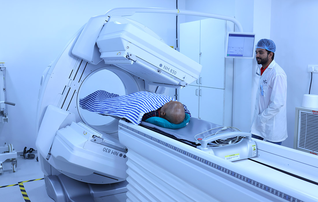
"BA" Cancer Centre is equipped with the G.E. dual head gamma camera machine with SPECT Capability.
Single Photon Emission Imaging/Tomography:
Single Photon Emission Imaging / Tomography is also a type of nuclear medicine imaging technique, which uses small amounts of radioactive material called radiotracers or radiopharmaceuticals to help diagnose and assess medical conditions.
Radiotracer like Technetium labelled pharmaceuticals (DTPA, DMSA, MIBI, MDP, HIDA etc.) are injected intravenously and imaging is performed under a gamma camera. Other radiopharmaceutical like I-131 is administered orally and the scanning is performed under a gamma camera using special collimator. The selection of the radiotracer depends on the medical condition to be imaged, as predetermined by national and international guidelines.
Few Common uses of Single Photon Emission Imaging/Tomography